

Time-of-Flight Photoelectron Emission Microscopy
Photoelectron Emission Microscopy (PEEM) is a surface sensitive tool to study topographic, elemental or chemical properties, electronic band structure or surface fields (electrical, magnetic) on a nanoscale. It is utilizing ultraviolet or soft X-ray excitation to liberate photoelectrons, whose spatial emission distribution or angular distribution can be imaged by a high resolution imaging electron microscope.
We are operating a dedicated electrostatic PEEM for ultrashort High Harmonic excitation pulses at high repetition rates. The microscope is equipped with two integrated electron energy filters, a Retarding Field high-pass filter as well a Time-of-Flight (ToF) spectrometer with Delay Line Detector anode, which allows for energy-filtered imaging and recording of kinetic energy spectra, when triggered by a pulsed excitation source.
By means of this scientific instrument we are studying localized surface plasmons and plasmon polaritons from metallic nanostructures on surfaces resolved on a spatial nanoscale. 4D microscopy is achieved in an optical-pump/soft X-ray-probe setup, with time resolution provided by the delay of the two ultrashort pulses, spectroscopic electron energy resolution provided by the ToF spectrometer and spatial resolution provided by the PEEM.
Our scientific goal is the characterization of collective electron motions and ultrafast charge transfers on attosecond (100-1000 as) temporal and nanometer (50-100 nm) spatial scale, a technology dubbed “ATTO-PEEM”.
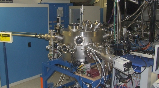
TOF-PEEM in UHV chamber for ultrafast plasmonic studies